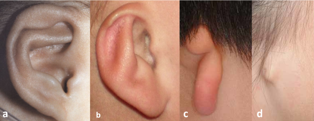
The ear reconstruction with autologous tissue, i.e. the reconstruction of the anatomy of the ear with cartilaginous tissue harvested from the patient, is a surgical procedure that is performed in case of partial or total absence of the outer ear due to a congenital malformation called microtia (Figure 1) or after burns. The surgery consists in the harvesting of the costal cartilage and subsequent sculpting and carving of the costal cartilage in order to obtain a framework that reproduces the mirrored shape and size of the contralateral healthy ear. This type of surgery is a real challenge for surgeons because obtaining an accurate anatomical reproduction of the ear can be extremely complicated due to the unique shape and size of this element.

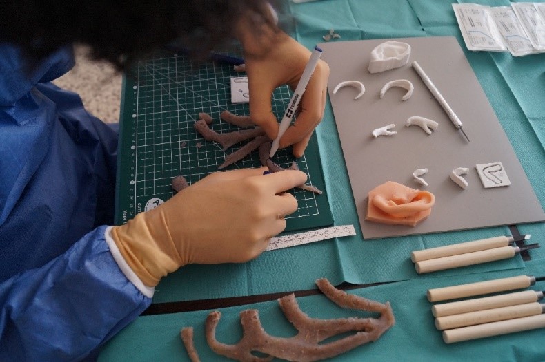
Operations require a high degree of manual skill and experience from the surgeon. In order to provide the clinician with a useful tool for reconstruction, the T3ddy team has designed custom cutting guides to actively assist the surgeon both in the pre-operative planning phase and during surgery (Figure 2).

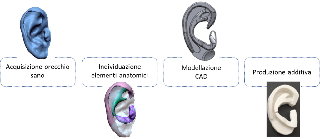
The realization and design of the surgical guides for each patient follows the workflow shown in Figure 3 and includes the acquisition of the healthy anatomy of the ear, the identification of the anatomical elements to be reconstructed, CAD modeling and realization with additive manufacturing technologies (3D printer) of the surgical guides.

The guides designed and manufactured by T3Ddy laboratory engineers in collaboration with the surgeons of Meyer Children’s Hospital represent a simplification of the ear anatomy while maintaining the main features of the anatomical element to be reproduced as shown in Figure 4.

The guides, printed with biocompatible materials, have been used in numerous surgeries at the Meyer Children’s Hospital and have considerably improved the aesthetic results of this surgery, as well as reducing intraoperative times.

